Diagnostic Ultrasound Findings in Gynaec lesions
Case Study Posted By : Dr. Manju Virmani
Description

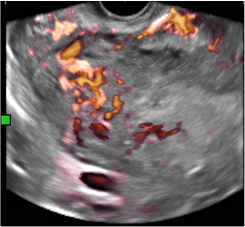
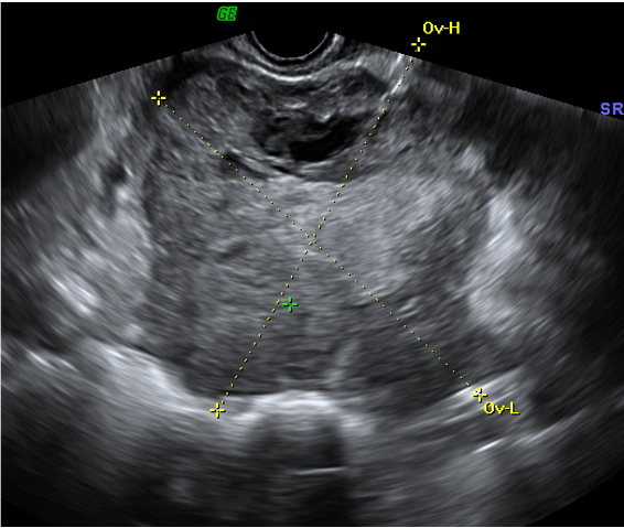
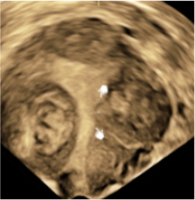
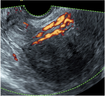
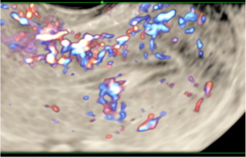
Ovarian malignancy
Irregular solid tumor (more than 80% solid)
presence of ascitis
largest diameter > 100mm
Strong blood flow color score 3 - 4
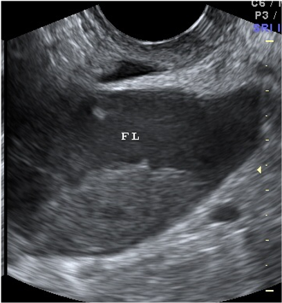
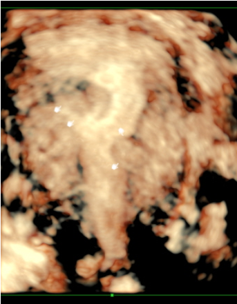
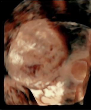
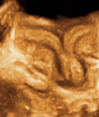
Mature Cystic Teratoma (Dermoid Cyst) Bilateral
most characteristic ultrasound features is white echogenic “ball” Rokitansky nodule or dermoid plug (corresponds to the sebum and hair content of the dermoid)
long and short thin linear strands demoid mesh – hair in fluid content
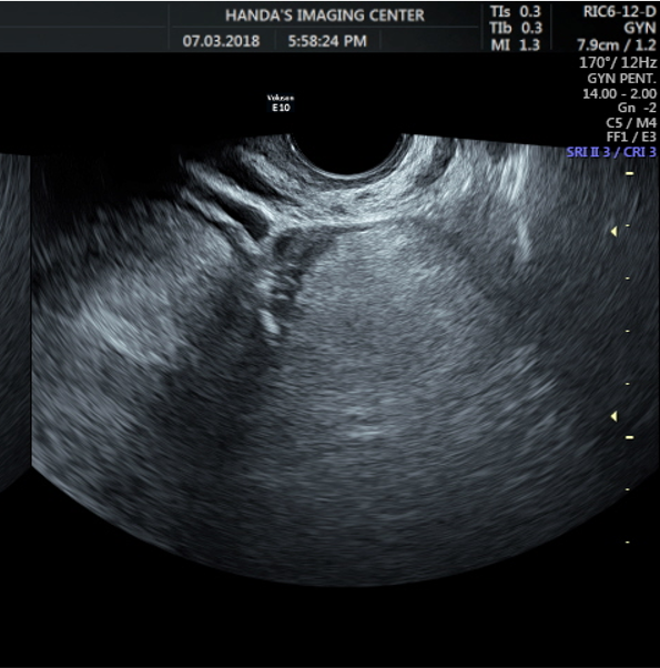
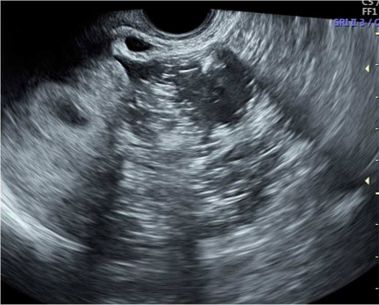
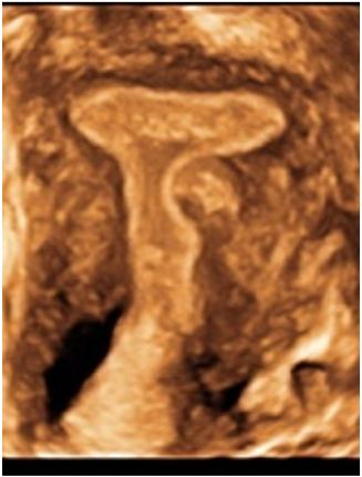
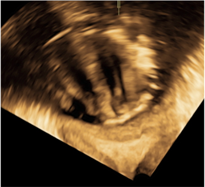
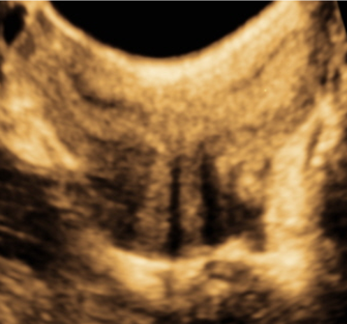
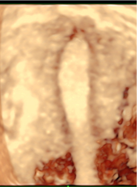
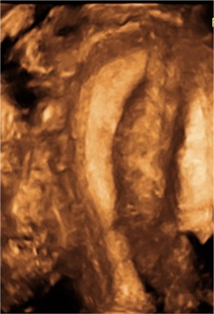
3D Hydrosalpinx 3D in inverse mode
Anechoic tubular structure in the adnexa separate from the ovary
C shaped tubular structure
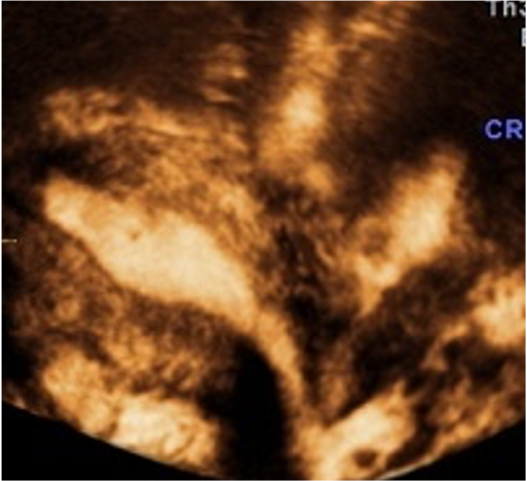
Incomplete Septa
0 – intra cavitary 1–submucosal. < 50% intramural 2-submucosal > 50% intramural. 3-100% intramural in contact endometrium
4 - intramural 5 – subserosal > 50 % intramural 6 – subserosal < 50 % intra mural. 7 - subserosal pedunculated
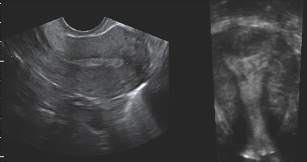
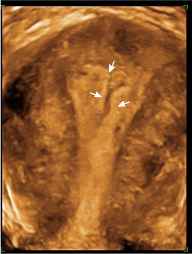
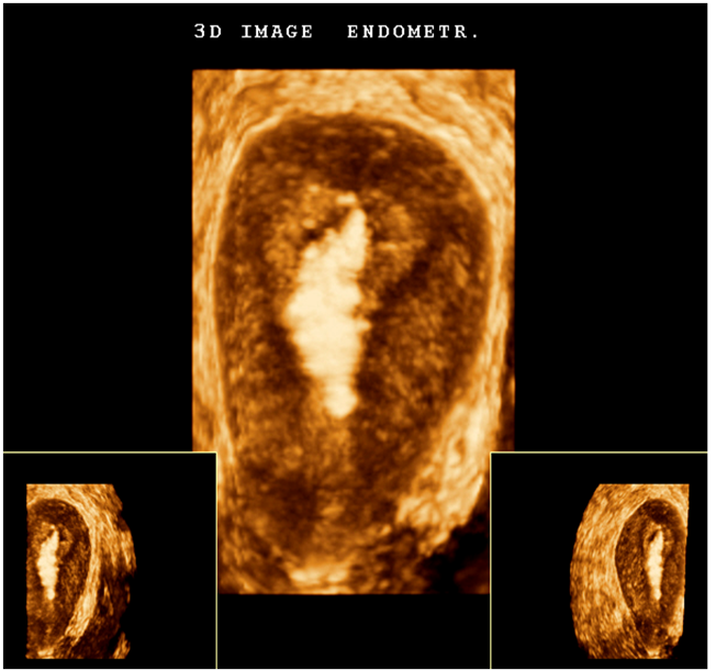
Polyps
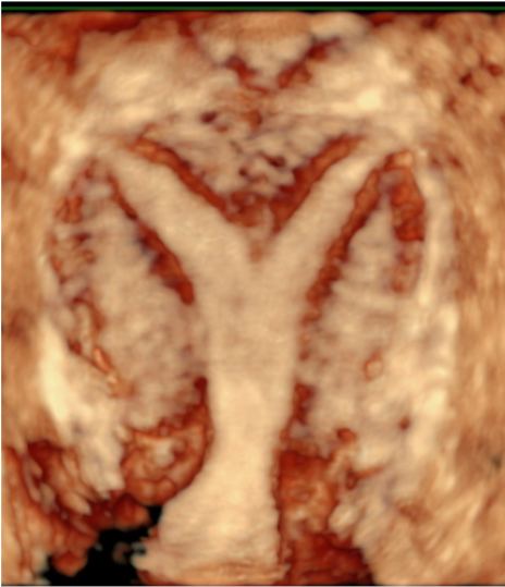
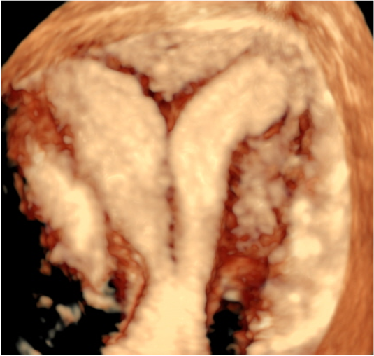
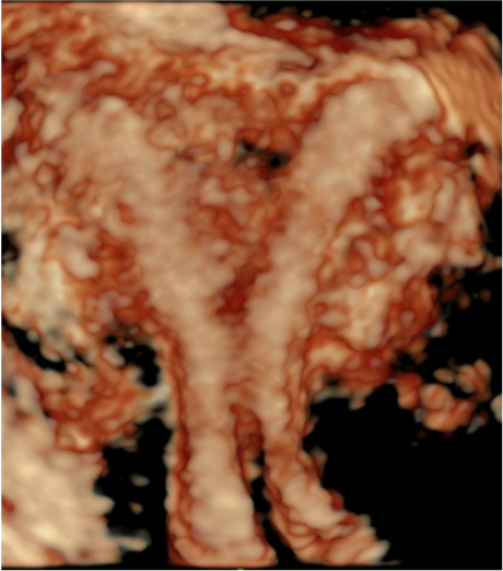
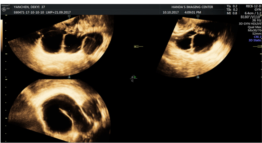
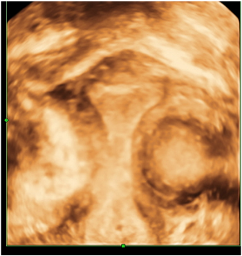
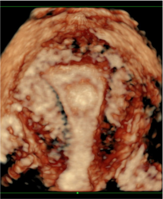
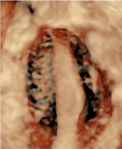
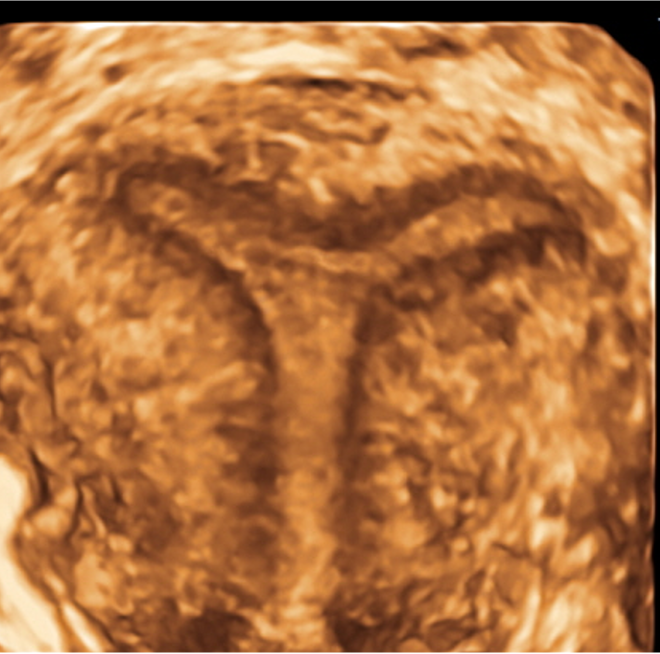
Partial Septate Complete Septate. Bicervical Septate Uterus
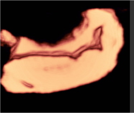
MDA
MDA Bicorporeal Uterus
Unicornuate Uterus: 20% MDA
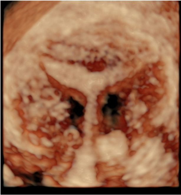
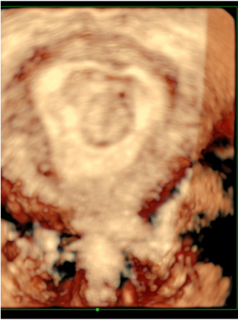
Dysmorphic T-shaped
Uterine adhesions
Retained calcified missed aborted tissue